|
click to home
Optical Coherence Tomography (OCT)
OCT is an imaging technique developed to image the various retinal layers and reconstruct 3-D retinal structure. It works on the principle of low coherence
interferometery where a light ray of low coherence (800-1300nm) is directed onto the eye and the back scattered light from the different retinal tissues
capture the information regarding the health of the retina. The OCT devices that use the above mechanism of capturing the tissue characteristics is called time
domain OCT. In order to capture the OCT scan of a region of retina, the OCT probe beam is adjusted to a point and a low coherence light is projected onto the
eye. The reflected light is plotted as a 1-D profile across depth known as A-scan. Similar operation is performed at different transverse locations known as Bscans.
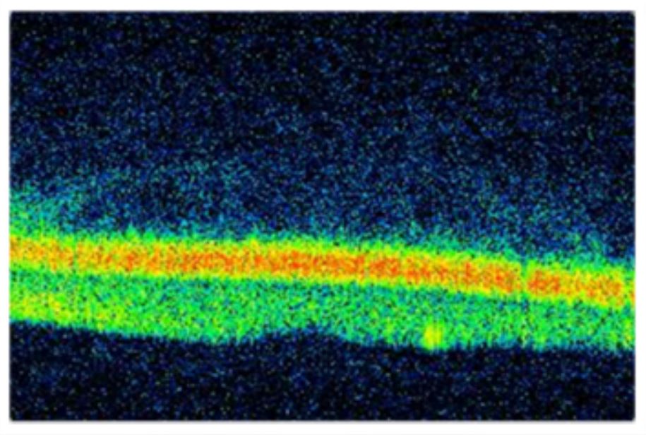
For better visualisation of the retinal layers, the B-scans are projected to a false color scale. Figure below shows a transverse (B-scan) of the macular region
of the retina obtained from OCT. The different colors in the figure show the optical characteristics of the tissue structure of the retina. A 3-D view of the
retinal tissue characteristics is obtained by a raster scan of the OCT probe across the retinal surface under study.
An estimate of the tissue density can be obtained by measuring the thickness of each layer. Also gaps or detachments in the layers can be studied. OCT is an
accepted medical imaging technique for diagnosis of diseases like diabetic retinopathy, glaucoma and age related macular degeneration.
 Fast acquisition of OCT scan leads to a better image capture as it prevents the movement of the eye by the patient. Therefore Fourier domain OCT machines
are preferred as they provide faster axial and transverse scans incomparison to the conventional time domain OCT devices.
Fast acquisition of OCT scan leads to a better image capture as it prevents the movement of the eye by the patient. Therefore Fourier domain OCT machines
are preferred as they provide faster axial and transverse scans incomparison to the conventional time domain OCT devices.
OCT has a very promising research future in both medical and engineering fields. In medical sector, reports obtained from the OCT can aid a physician to decide
the progress of retinal diseases in spite of performing numerous invasive tests that cause a lot of discomfort to the patients. The images obtained by the OCT
machine are highly degraded by speckle noise and do not show clear and sharp boundaries of the different layers. Image processing tools can be devised to enhance
the image quality opening doorway for medical research for signal processing engineers.
Vineeta Das
Research: Biomedical Image Processing (EMST LAB)
Email: vineetadas@iitg.ernet.in
|
 |
|