Welcome to Bosanta's homepage

« Previous Page
My Teaching Academics My Research Research Group Publications Openings My family

Confocal and super-resolution Microscopy

Laser scanning confocal microscope can provide superior axial resolution relative to widefield microscope. Since the images obtained with a confocal microcope are devoid of any out of focus blur, the technique is very popular amongst biologist to image deep into a tissue without any physical insertion into the sample. Figure 1 shows a lens "L" focusing the light emitted by two point sources (red one and the blue one). The red source is considered to be in the object plane (or the sample plane) and the light from it are focused at a point where a pinhole "PH" is positioned. It is now seen that the pinhole allows more light from the red source to reach the photodetector than the light from the blue source which is behind the object plane.

Figure 1: The principle of obtaining superior axial resolution.

Similarly it is easily seen that the light from a source ahead of the object plane are also mostly blocked by the pinhole. The principle depicted in figure 1 is used in a confocal microscope to block the out of focus light from reaching the image plane. In order to form an image of a 2D area, a focused laser beam scans the target. For each position of the laser beam, light scattered or emitted by the target is descanned by the scanner and focused onto the pinhole. Signal from the photodetector is sent to a computer, which at the end of the scan forms an electronic image of the target area. Using an illumnation laser beam of wavelength λ the lateral and axial resolutions achievable with a confocal microscope are λ/2 and λ respectively.

The performance of a confocal microscope may be compromised due to the presence aberrations in the illumnation beam or some times due to the aberrations introduced by the target being imaged. By incorporating a programmable optical element in the laser beam path of a confocal microscope it is possible to have an illumination beam with a reconfigurable wavefront. Thus knowing the aberrations being introduced, it is possible with the programmable element to get a detection path that is free from aberrations. Figure 2 shows the focus spots in the sample plane of a confocal microscope with a helical wavefront illumination beam. Due to the abberations present in the laser beam the focus spot (left) is not a perfect doughnut, while the focus spot (right) is closer to a doughnut as the illumination beam has been partially corrected for aberrations using the programmable element.

In addition to correcting for aberrations the programmable diffractive element in the illumination beam path of the confocal microscope can be used to modulate the polarization profile. Thus one can do confocal imaging using various vector beams instead of the conventional scalar beam. We have developed a confocal system where the illumination beam can be periodically or aperiodically switched between different polarization states of the illumination beam. In Figure 3 we show confocal images of liquid crystal pixels in the ON or OFF state which correspond to orthogonal molecular orientaitons when the illumination beam is switched between X and Y polarizations after every line scanned. It is noticed that the same molecule with different orientations responds to X and Y polarizations in a different manner. This demonstrates the dependence of the spotanenous emission probability with the angle between the excitation field and the electric dipole associated with the moeculecule. More details about a confocal microscope with vecctor beam illumoination is available here.

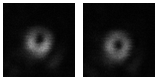
Figure 2: The illumination beam spots with a helical wavefront in the presence of aberration (left) and after partial correction of aberrations (right).

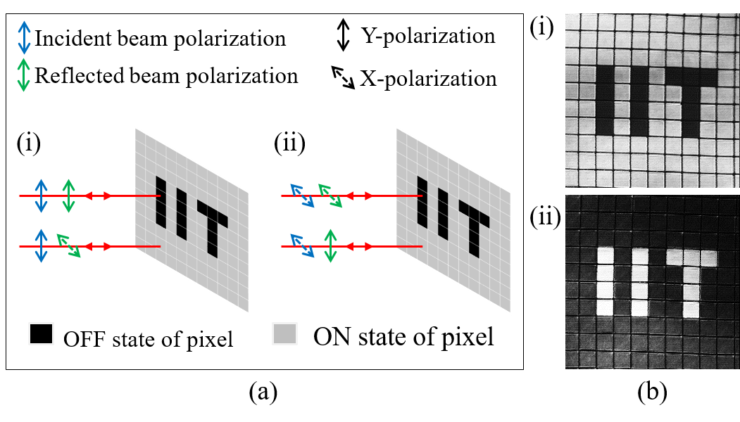
Figure 3: Schematic showing the change in polarization of the illumination beam by the ON/OFF pixels (left) and confocal images of liquid crystal pixels arranged as the "IIT" (right).

A confocal microscope can not resolve details in an object which are much smaller than the wavelength of light due to the diffraction limit. A microscope that can display finer details beyond the diffraction limit is called a super-resolution microscope. There are two successful optical super-resolution techniques for imaging of fluorescent molecules which were also awared the Nobel prize in the year 2014. One of these techniques uses the stimulated emission depletion phenomenon in an state of the art optical arrangement.

In the other technique the diffraction barrier is overcome by making use of the stochastic nature of the fluorescence emmission. We have recently developed one such imaging system called Stochastic Optical Reconstruction Microscope (STORM) in our laboratory. Figure 4 demonstrates how images using STORM can show finer details upto a resolution of 30 nm in contrast with a widefield microscope.

Figure 4: Images of cells using (left) a wide microscope and (right) a super-resolution STORM microscope.